
 TR
TR
 EN
EN


ABSTRACT
Objective
The main purpose of this study was to investigate serum angiotensin-converting enzyme (ACE) activity levels in pediatric patients diagnosed with community-acquired pneumonia (CAP) and healthy controls and to determine the link between serum ACE activity and white blood cells (WBC), neutrophil (NEU) and C-reactive protein (CRP) levels.
Materials and Methods
Children aged between 1 month-13 years were included in the study. The diagnosis of CAP was based on clinical symptoms- such as cough, sputum, fever or chills, tachycardia, tachypnea, chest retractions, pulmonary auscultation findings- and radiographic evidence of alveolar infiltration or consolidation. The CAP group was further classified as bacterial (n= 50) or viral (n= 31) and compared with healthy controls (n= 54). Serum ACE activity, WBC, NEU, and CRP levels were measured
Results
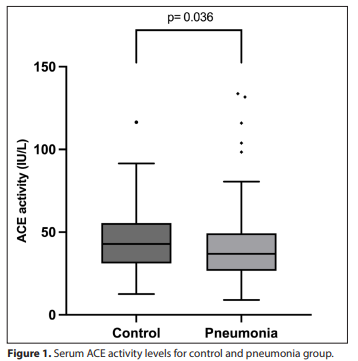
There were significant differences between the patients with CAP and healthy controls in serum ACE activity, WBC count, NEU count, and CRP level (p= 0.036, <0.001, <0.001, and 0.022, respectively). However, no significant difference in serum ACE activity was found among the three groups. No correlation was observed between serum ACE activity and age, WBC, NEU, or CRP levels. The area under the ROC curve for serum ACE activity in predicting CAP was 0.60 (95% Confidence interval: 0.51–0.70, p= 0.035).
Conclusion
Serum ACE activity can distinguish CAP patients from healthy controls. However, it is not an effective marker for differentiating between viral and bacterial pneumonia. In addition, each laboratory should establish its own reference interval for serum ACE activity based on the analytical method employed and the relevant age groups.
KEYWORDS
Angiotensin-converting enzyme, community-acquired pneumonia, children, differential diagnosis
INTRODUCTION
The role of serum angiotensin-converting enzyme (ACE) in the regulation of blood pressure and electrolyte homeostasis is well established. Although ACE is found in many tissues and cell types, it is predominantly expressed on the surface of epithelial cells in the lungs, intestines, kidneys, and blood vessels (1,2). When blood pressure decreases, the renin-angiotensin-aldosterone system is activated, leading to the secretion of renin from the kidneys. Renin catalyzes to conversion of angiotensinogen to angiotensin-I. Subsequently, angiotensin I is converted to its highly active form, angiotensin II, by the action of ACE. Angiotensin II induces vasoconstriction and stimulates aldosterone release, resulting in an increase in blood pressure (3). Following this reaction, ACE expressed in the endothelium is released into the circulation. Community-acquired pneumonia (CAP) is one of the most common diseases of the childhood worldwide and is characterized by acute inflammation of the lung parenchyma in response to infectious agents such as bacteria and viruses (4- 6). CAP presents clinically with high fever, signs of respiratory distress, laboratory evidence of infection, and parenchymal involvement, which are identified through physical examination and/or chest radiography (6,7). Numerous studies have examined serum ACE levels in lung diseases such as sarcoidosis, pneumonia, and acute respiratory distress syndrome (8-12). Three primary hypotheses have been proposed to explain the decrease in serum ACE activity during pneumonia (9). The first suggests that the inflamed pulmonary vascular endothelium produces and releases lower amounts of the enzyme. The second hypothesis posits that the demand for angiotensin II increases during pneumonia in order to maintain pulmonary homeostasis. The third hypothesis involves the presence of ACE-inhibiting compounds—such as nitric oxide, nitric oxide-releasing compounds, or reactive oxygen species—that may circulate during pneumonia. In this study, we aimed to evaluate the utility of serum ACE activity in distinguishing pediatric patients diagnosed with CAP from healthy children, using routine examinations and tests performed at hospital admission. Additionally, we sought to investigate the role of serum ACE activity in the differential diagnosis of CAP. Secondary objectives included determining the cut-off value for serum ACE activity and assessing its correlation with white blood cell (WBC) count, neutrophil (NEU) count, and C-reactive protein (CRP) in the context of CAP diagnosis.
MATERIALS AND METHODS
Study design and population This study was conducted at XXX Training and Research Hospital, YYY, Türkiye, with the approval of the local Ethics Committee (approval no: xxx) and in accordance with the principles of the Declaration of Helsinki. Written informed consent was obtained from all patients or their legal guardians. The study population consisted of individuals of white-Caucasian ethnicity. This was a prospective observational study conducted between October 2021 and March 2022 in the Department of Pediatrics. Children aged 1 month to 13 years who were hospitalized with symptoms suggestive of CAP were included, along with a control group of healthy children presenting to the outpatient clinic for routine health evaluations. The diagnosis of CAP was established based on previously published guidelines, considering clinical symptoms such as cough, sputum production, fever or chills, tachycardia, tachypnea, chest retractions, pulmonary auscultation findings, and radiographic evidence of alveolar infiltration or consolidation consistent with CAP (7,13,14). All X-ray images were interpreted by a radiologist blinded to the children’s diagnoses. The radiologist classified the X-ray images as compatible with bacterial/viral pneumonia or both conditions. Based on these findings, patients were categorized as having either viral or bacterial pneumonia, and appropriate treatment was initiated accordingly. According to the Turkish Thoracic Society Consensus Report on the Diagnosis and Treatment of CAP in Children, diagnostic investigations to identify the causative pathogen are recommended only for hospitalized patients (15). Since all patients in our study were outpatients who presented to the pediatric clinic and were followed up on an outpatient basis, neither sputum cultures nor respiratory viral PCR panels were performed. Subjects with chronic diseases, malignancies, regular medication use (ACE inhibitors, angiotensin II receptor blockers or immunosuppressive therapy including systemic corticosteroids), or a history of hospitalization within the past 30 days were excluded from the studySample collection At the time of admission, 2 mL of venous blood was routinely collected into K2EDTA tubes (BD Diagnostics, Franklin Lakes, NY, USA) for complete blood count (CBC) analysis and into gel-separated serum tubes (Vacutainer® SST™ II Plus, BD Diagnostics, Franklin Lakes, NY, USA) for biochemical examination. Serum samples were obtained by centrifugation at 2500 g for 15 minutes at 4 °C within one hour of venipuncture. Residual serum from the biochemistry analysis was aliquoted into microtubes for measurement of serum ACE activity. Hemolytic, lipemic, and icteric samples were excluded based on LIH index analysis. One study demonstrated that serum ACE activity remains stable for only up to one week when stored at -20 °C (16). Other studies have reported that serum or plasma ACE activity generally remains stable for several months or years at -20 °C; however, freezing and thawing serum or storing it at -70 °C can increase ACE activity by approximately 15% (17,18). Therefore, in this study, samples were stored at -20 °C for a maximum of one week and were thawed and analyzed. Analytical measurements CBC and biochemistry analyses were performed on the day of admission. WBC and NEU counts were measured based on flow cytometry method using the semi-conductor laser beam on the Sysmex XN 3000 instrument (Sysmex Co., Kobe, Japan). CRP levels were measured by the particle-enhanced immunoturbidimetric method on Roche Cobas 8000 autoanalyzer (Roche Diagnostics, Indianapolis, IN, U.S.A.). Serum ACE activity was measured using Roche Cobas c501autoanalyzer (Roche Diagnostics, Indianapolis, IN, U.S.A.) with ACE reagents supplied by BEN Biochemical Enterprise (Milan, Italy). ACE activity was determined by the kinetic colorimetric method using FAPGG (N- (3-(2-furyl)acryloyl)-L-phenylalanylglycylglycine) as the substrate. The decrease in absorbance of FAPGG at 340 nm and 37 °C per unit time is proportional to the ACE activity in the sample. Interferences for triglyceride, hemoglobin and bilirubin are constant for up to 1000 mg/dL, 300 mg/dL and 20 mg/dL, respectively. Calibration and internal quality control studies for serum ACE activity were performed prior to analysis. According to the manufacturer, limit of detection is 2.6 IU/L and method linearity is up to 150 IU/L. The manufacturer’s reference interval is 8–-2 IU/L. The intra-assay coefficient of variation (CV) is 1.3%, and the inter-assay CV is 2%. Statistical analysis Data were analyzed using the Statistical Package for the Social Sciences (SPSS) version 29.0 (IBM Corp., Armonk, NY, USA) and GraphPad Prism (version 10; GraphPad Software, Boston, MA, USA). Kolmogorov- Smirnov test was used to assess distribution of clinical data. As all variables were non-normally distributed, results are presented as median and range. The Mann-Whitney U test was used to compare medians of WBC, NEU, serum ACE activity, CRP levels and age between patients with pneumonia and control group. The Kruskal-Wallis test was employed to compare these parameters among the bacterial pneumonia, viral pneumonia, and control groups. Pairwise comparisons were performed using the Mann-Whitney U test with Bonferroni correction to adjust for multiple comparisons. Spearman correlation analysis was conducted to examine the relationship between serum ACE activity and other parameters. For multivariate analysis, logistic regression analysis was performed to identify independent predictors of pneumonia diagnosis. Receiver operating characteristic (ROC) analysis was conducted to evaluate the sensitivity and specificity of serum ACE activity in predicting pneumonia.
RESULTS
A total of 140 children (72 boys and 68 girls) were initially enrolled in the study. However, four boys and one girl were excluded due to not meeting the inclusion criteria or insufficient blood sample availability. Consequently, the final analysis included 135 children (68 boys and 67 girls), aged 1 month to 13 years (median age: 2 years). Among these, 54 (40%) children were classified as healthy controls, while 81 (60%) children were diagnosed with pneumonia, comprising 31 with viral pneumonia (23%) and 50 with bacterial pneumonia (37%). Mean body temperature of the participants was 38.2 °C (standard deviation= 1.0; range: 35.6-41.0 °C). Demographic and laboratory data of the patients diagnosed with pneumonia and the control group are presented in Table 1. There were significant differences between the pneumonia and control groups in terms of serum ACE activity, WBC count, NEU count, and CRP level (p= 0.036, <0.001, <0.001, and 0.022, respectively). WBC count, NEU count, and CRP level were significantly higher in patients with pneumonia, whereas serum ACE activity was significantly lower in this group (Figure 1). There was no significant difference in age between the two groups. Additionally, when age, WBC, NEU, CRP level, and serum ACE activity were compared by sex, no significant differences were observed between boys and girls. Patients diagnosed with pneumonia were further classified as having either bacterial or viral pneumonia and compared with the control group. There was no significant difference in serum ACE activity among the three groups (Figure 2). Analysis of WBC count, NEU count, and CRP levels revealed significant differences between the bacterial and viral pneumonia groups, as well as between the bacterial pneumonia and control groups; however, no significant difference was observed between the viral pneumonia and control groups (Table 1). Correlations between serum ACE activity and age, WBC count, NEU count, and CRP level were evaluated separately, and no significant correlations were identified. In contrast, a very strong positive and significant correlation was observed 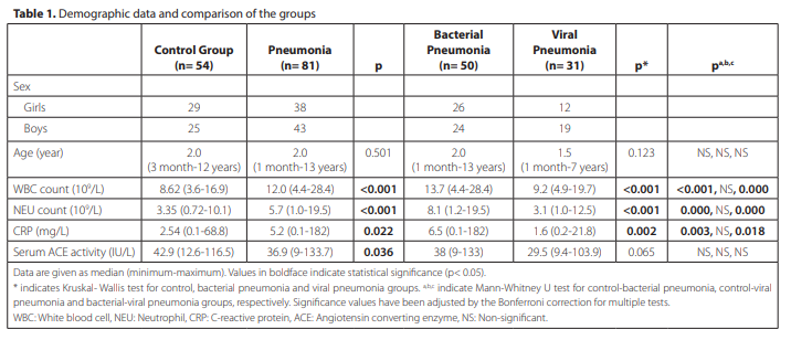
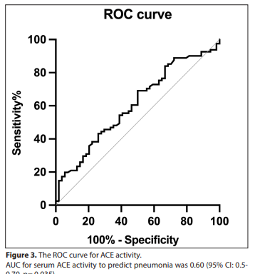
between WBC and NEU counts (r= 0.81, p< 0.001). Weak but significant correlations were found between age and NEU count (r= 0.30, p< 0.001), CRP and WBC count (r= 0.25, p< 0.001), and CRP and NEU count (r= 0.34, p< 0.001). Detailed correlation data are presented in Table 2. Area under the ROC curve (AUC) for serum ACE activity to predict pneumonia was 0.60 [95% confidence interval (CI): 0.51-0.70, p= 0.035] (Figure 3). The optimal cut-off value was determined as 44.4 IU/L, with a sensitivity of 69.1% and a specificity of 50.9%. The positive likelihood ratio was calculated as 1.41, and the negative likelihood ratio was 0.61.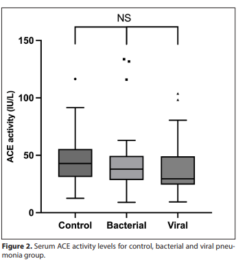
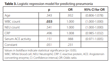
Logistic regression analysis was performed to assess the association of age, WBC count, NEU count, CRP, and serum ACE activity with the diagnosis of pneumonia. WBC count demonstrated a statistically significant association (p= 0.033), but the odds ratio was 1. None of the variables were identified as significant independent predictors. The multivariate model predicted pneumonia with an accuracy of 66.7%, whereas serum ACE activity alone showed a predictive accuracy of 58.5%. Detailed results of the logistic regression analysis are presented in Table 3.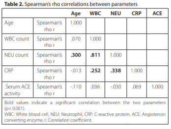
DISCUSSION
Pneumonia is one of the most common diseases in the childhood. In this study, we explored the potential role of serum ACE activity as a biomarker to distinguish children with CAP from healthy peers and to differentiate between bacterial and viral etiologies. Serum ACE activity was found to be significantly reduced in patients with pneumonia compared to healthy controls, consistent with the findings of previous studies (12,19). However, its ability to independently discriminate disease status was limited. Although ACE activity was also lower in viral pneumonia than in bacterial pneumonia, this difference did not reach statistical significance. Notably, when ACE activity was evaluated in combination with inflammatory markers such as WBC count, NEU count, and CRP, an improvement in overall diagnostic accuracy was observed. This suggests that ACE may have potential as a complementary marker within a multiparametric diagnostic approach, rather than serving as a standalone indicator. Only a limited number of studies have evaluated the relationship between serum ACE activity levels and pneumonia, and these have primarily involved adult patients (9,12,20,21). Although several studies have investigated serum ACE activity in pediatric populations, none have specifically focused on pediatric patients with pneumonia (22-24). In the study by van de Gerde et al., a significant decrease in serum ACE activity was observed during episodes of pneumonia, with levels returning to the reference range during recovery (21). The reduction in ACE activity was similar between the bacterial and viral pneumonia groups, with mean serum ACE activities at admission for both groups reported as 29 IU/L. In our study, serum ACE activity at admission was 29.5 IU/L in patients with viral pneumonia and 38 IU/L in those with bacterial pneumonia. Although serum ACE activity was lower in the viral pneumonia group than in the bacterial pneumonia group, the difference was not statistically significant. However, it should be noted that the study by van de Gerde et al. was conducted in adult patients, which limits direct comparability with our pediatric population. In contrast, the study by Abouzeid et al. involving pediatric CAP patients reported that the mean ACE activity in healthy controls and CAP patients was significantly different, at 58 ± 12.6 IU/L and 36.7 ± 9.4 IU/L, respectively. These findings are consistent with both the results and the age range of subjects in our study (19). In studies involving adult patients with SARS-CoV-2 pneumonia, no significant difference in serum ACE activity was observed between patients and healthy individuals (25,26). Guler et al. have reported median serum ACE activities of 38.0 IU/L in patients and 32.0 IU/L in controls (25). On the other hand, Henry et al. have found median values of 41.1 IU/L in patients and 42.9 IU/L in controls (26). In our study, median serum ACE activities for patients with viral pneumonia and healthy controls were 29.5 IU/L and 42.9 IU/L, respectively. Consistent with these studies, we did not observe a significant difference between the two groups; however, the serum ACE activity in our patients with viral pneumonia was lower than that reported in the aforementioned studies. Children and adolescents have higher serum ACE activity compared to adults (23,24). Using a FAPGG-based enzymatic activity assay, one study reported plasma ACE levels ranging from 13 to 100 IU/L in children (6 months to 17 years), and from 9 to 67 IU/L in adults (27). In another study conducted by Rodriguez et al., serum ACE activity was found to be 46.7 ± 11.93 IU/L in children (0 month to 15 years) and 32.1 ± 8.53 IU/L in adults, using a different method (23). Bénéteau-Burnat et al. compiled adult ACE reference intervals (RIs), and recommended standardizing measurement conditions- specifically, using FAPGG as the substrate at a concentration of 0.8-1.0 mmol/L, a wavelength of 340 nm and HEPES-pH 8.2 buffer, with appropriate differential absorptivity established for each device (17). Similarly, in our measurement, the wavelength used was 340 nm and the FAPGG substrate concentration specified in the kit insert >0.25 mmol/L. Muller summarized studies on serum ACE activities measured using the FAPGG substrate, highlighting the variability of RIs (28). This variability has been attributed to the influence of ACE insertion (I)/deletion (D) genetic polymorphism and differences in testing protocols (28,29). As a result, there are various methods and reagents for measuring ACE activity, and applying a single RI instead of ACE I/D specific RIs may reduce the precision of ACE activity interpretation (29-32). Consequently, RIs differ among studies, and standardization for this test has not been achieved (17,29). Therefore, each laboratory should establish its own RIs for serum ACE activity. This study has several limitations. First, blood and/or sputum cultures and respiratory viral PCR panel were not used to distinguish between bacterial and viral pneumonia, as these diagnostic tests are not required for outpatients; diagnoses were made based on clinical and radiological findings evaluated by a pediatrician. In addition, we did not include other relevant biomarkers such as angiotensin II, ACE protein, or procalcitonin. Second, data were limited to samples collected at the time of hospital admission, focusing solely on differential diagnosis. Clinical course and prognostic outcomes were not assessed. Follow-up measurements of serum ACE activity, WBC count, NEU count, and CRP levels after treatment were also unavailable due to sample limitations in pediatric patients. Third, participants were not genotyped for the ACE insertion/deletion polymorphism, which may influence serum ACE activity. Finally, median age of the participants was two years, which limits the generalizability of our findings to older children. Future studies including a broader pediatric age range are needed to validate our results across different developmental stages.
CONCLUSION
In conclusion, our study is notable as one of the few to evaluate serum ACE activity in pediatric patients with pneumonia. Although serum ACE activity was significantly reduced in patients with pneumonia compared to healthy controls, it was not sufficient to distinguish between bacterial and viral pneumonia. Additionally, since serum ACE activity differs from adult values and can vary according to the method used and age groups, each laboratory should establish its own reference interval.